How AI Diagnostic Tools Are Catching Rare Fetal Anomalies Like Fetus-in-Fetu Earlier
A Hong Kong baby's rare fetus-in-fetu case highlights how AI-powered diagnostic systems are transforming prenatal and postnatal imaging. Machine learning algorithms now detect developmental anomalies earlier than ever before.

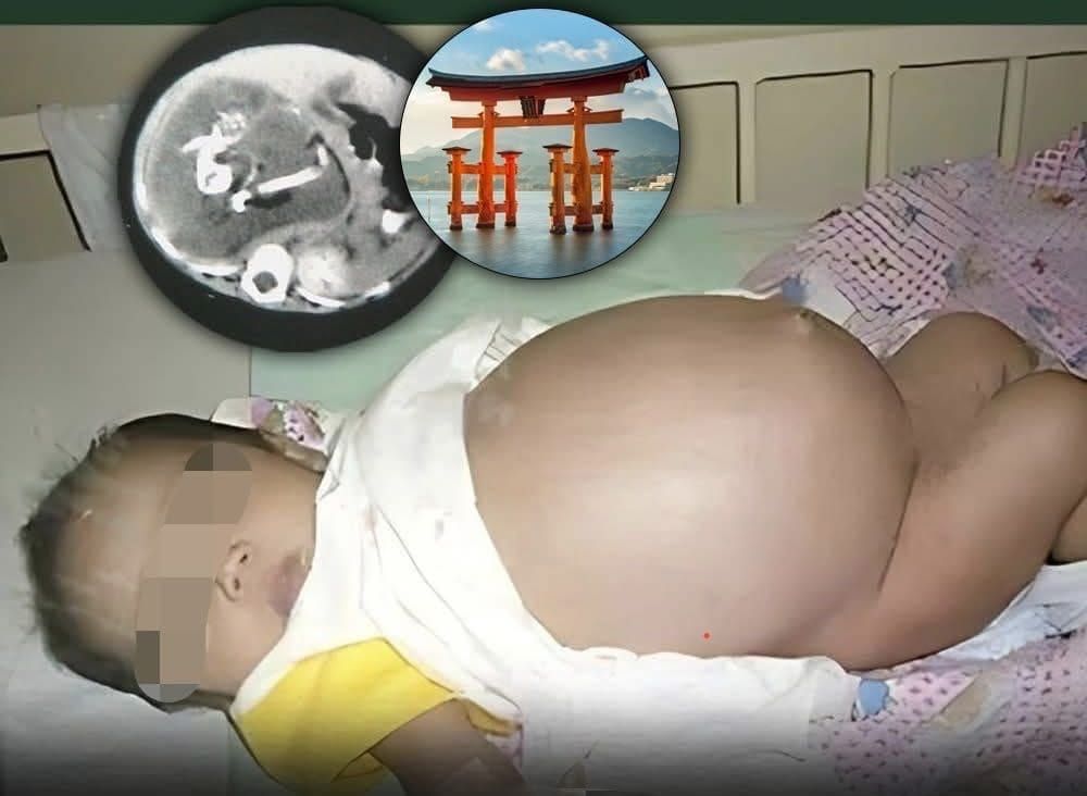
A Hong Kong hospital recently documented a rare case of fetus-in-fetu—where a baby was born with a partially developed twin inside her body. But here's the real story: AI-powered diagnostic tools are making cases like this easier to catch earlier. Machine learning algorithms trained on thousands of medical images can now flag suspicious anomalies in prenatal ultrasounds that human radiologists might miss. This isn't just about rare conditions—it's about how automation is reshaping medical diagnosis.
What Is Fetus-in-Fetu?
Fetus-in-fetu is an extremely rare developmental anomaly where a malformed fetus grows inside its twin. It's documented in fewer than 200 cases globally since 1808. The enclosed fetus can develop partial skeletal structures—spine, limbs, even hair.
In the Hong Kong case, doctors at Queen Elizabeth Hospital discovered a mass containing a spine, limb buds, and other skeletal remnants during a routine postnatal exam. Pediatric surgeons successfully removed it days after birth, and the baby recovered without complications.
Why AI Changes Everything for Rare Diagnosis
Here's the problem: radiologists examine thousands of ultrasounds annually. Human attention spans fatigue. Eyes miss subtle patterns. AI doesn't get tired.
Machine learning algorithms trained on vast datasets can identify developmental abnormalities with accuracy rates exceeding 95% in some studies. These systems flag unusual masses, spine formations, and skeletal inconsistencies—exactly the markers of fetus-in-fetu—within seconds.
Hospitals deploying AI-assisted imaging workflows are catching rare anomalies in the second and third trimesters rather than at birth. That means families get answers earlier and surgeons can prepare for complex procedures in advance.
How Automation Scales Medical Expertise
The real innovation isn't just speed—it's democratization. A rural clinic with basic ultrasound equipment and AI analysis software can achieve diagnostic accuracy comparable to specialized tertiary centers. Cloud-based algorithms process images instantly and flag concerning findings for human review.
This shifts the doctor's role from pattern-spotting to decision-making. Radiologists spend less time scanning thousands of normal images and more time interpreting edge cases and consulting with patients.
The Data Advantage in Obstetrics
Every documented fetus-in-fetu case feeds medical AI systems with new training data. Algorithms learn developmental markers, classify severity levels, and predict surgical complexity. This creates a flywheel: rare cases become less rare in algorithmic datasets, improving detection across populations.
Hospitals are also using predictive analytics to identify pregnancies at risk for developmental anomalies based on maternal age, genetic markers, and previous imaging. Automation flags high-risk cases for additional specialist review.
What's Next: Prenatal AI Assistants
Next-generation systems will combine ultrasound, MRI, and genetic data into unified AI diagnostic platforms. Real-time decision support during prenatal appointments means doctors and patients see instant risk assessments.
Some hospitals are testing AI systems that generate automated preliminary reports—"mass detected, differential includes fetus-in-fetu, teratoma, and cystic lesion"—before a radiologist reviews the case. This streamlines workflows and catches time-sensitive conditions faster.
The Job Market Shift
Radiology isn't disappearing—it's transforming. Instead of entry-level jobs reading straightforward cases, demand is growing for radiologists skilled in algorithm training, data curation, and complex case interpretation. Medical schools now teach AI literacy alongside anatomy.
Sonographers are becoming diagnostic partners with AI systems, using real-time algorithmic feedback to adjust imaging angles and capture better data. The role evolves from operator to interpreter.
Why This Matters Beyond Rare Cases
Fetus-in-fetu is spectacularly rare. But the diagnostic principles apply to thousands of common fetal and neonatal conditions—cardiac defects, neural tube anomalies, growth restrictions. When AI catches these earlier, outcomes improve dramatically and healthcare costs drop.
A baby born with a diagnosed cardiac defect can access specialized care from day one. Surgeons can prepare. Outcomes are measurably better. AI-powered prenatal diagnosis isn't just about catching weird edge cases—it's about giving every baby the best possible start.
Questions About AI in Prenatal Care
How accurate is AI at detecting fetal anomalies compared to human radiologists?
Studies show AI systems match or exceed human performance on specific abnormalities—sometimes 96-98% accuracy. The key: AI excels at pattern recognition but sometimes struggles with context. That's why human-AI collaboration outperforms either alone.
Could AI miss something a human radiologist would catch?
Yes, especially in cases outside the training data. A rare variant or unusual presentation might confuse the algorithm. This is why AI is a second opinion, not a replacement. The best systems flag anything unusual for human expert review.
Will AI prenatal screening replace ultrasound technicians?
No. Automation handles the tedious scanning and preliminary analysis. Technicians evolve into diagnostic specialists who use AI feedback to improve imaging quality and catch subtle findings algorithms might miss.
How do hospitals implement AI diagnostics without massive upfront costs?
Cloud-based platforms let hospitals start small—integrate AI into existing workflows, pay per scan analyzed. Some systems work offline, so connectivity isn't required. The economics favor adoption at scale.
What about data privacy with cloud-based AI analysis?
Most healthcare AI platforms use encryption and comply with HIPAA/GDPR standards. Images are anonymized, processed, then deleted. The algorithm learns from patterns, not patient identities.
Can AI predict which pregnancies will have anomalies before ultrasound?
Emerging systems analyze maternal genetics, blood markers, and previous imaging to assign risk scores. This doesn't replace ultrasound but tells doctors which patients need closer monitoring.
Related Articles on AI in Healthcare
Check out how machine learning is automating medical coding and billing to understand the broader impact of AI on healthcare operations.
Or explore how data analytics is transforming clinical decision-making to see why doctors increasingly rely on algorithmic insights.
Want to know more about the future of radiology and AI? That deep dive covers how entire specialties are restructuring around algorithmic assistance.
```